Hand Reconstruction Surgery

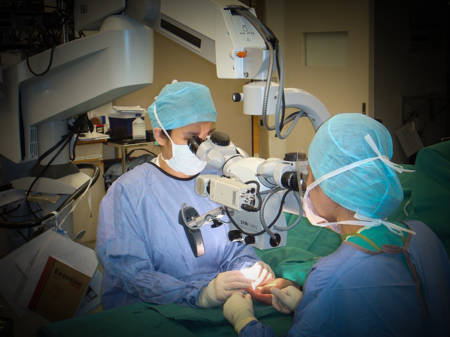
Microsurgery (surgery under magnification)

Microsurgery is performed using operating microscopes with high magnification (8-40x). This allows us to appreciate the anatomy of small but critical structures like nerves and blood vessels of the fingers or the limbs so that we can repair them to re-establish function. Fine sutures and micro-needles with special micro-instruments allow this surgery to be performed. The surgeon has to be specially trained in reconstructive microsurgery.

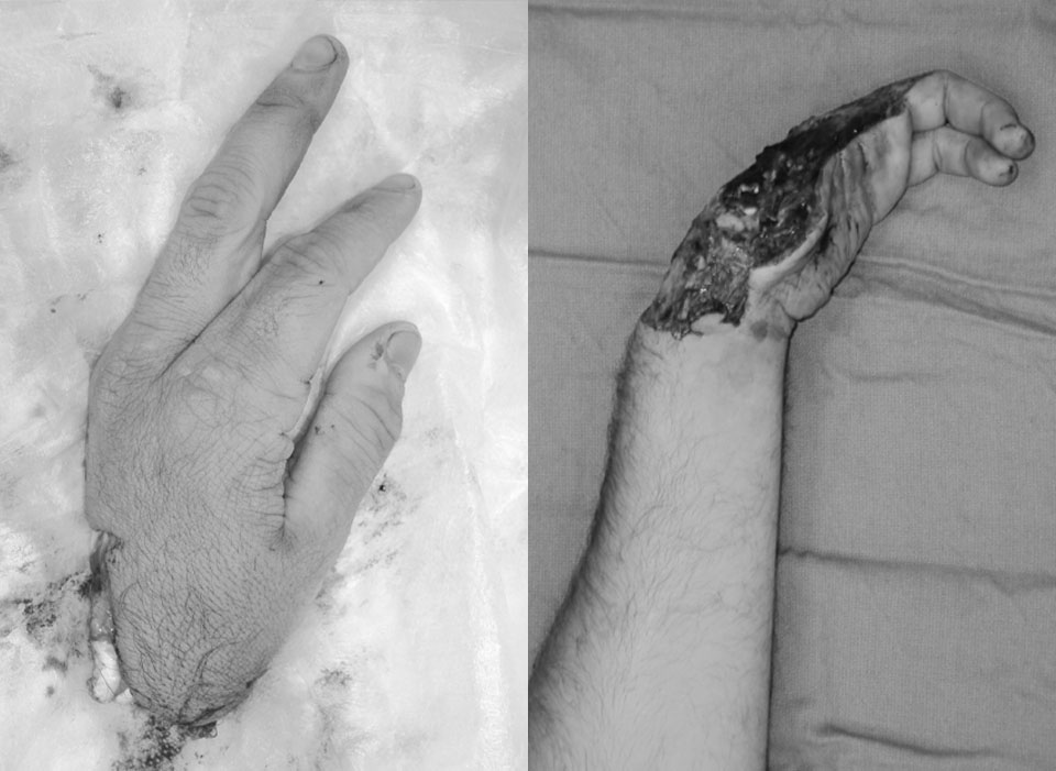
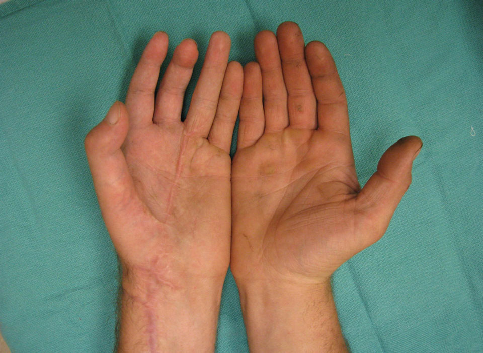
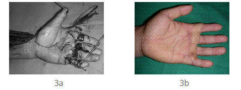
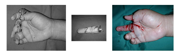
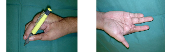
In severe trauma involving limb amputation, the only salvage is to re-establish blood flow to the limb by repairing the artery (inflow vessel), veins (outflow vessels) and the nerves. Microsurgery is critical for replantation to be successful.

Fig1. Microsurgeons at work

Fig1. Microsurgeons at work

Fig 2: Re-plantation of part of a hand which was amputated.

This advanced surgical skill is also used to salvage limbs after resection surgery for cancer, infection or after tissue loss following trauma. Free tissue transfer refers to the transfer of tissue from one part of the body to reconstruct critical tissue losses in another part. To ensure viability of the transfer, successful microsurgical anastomosis of critical arteries and veins are vital.

Fig 3a. Critical defects over multiple digits resurfaced with a flap from the forearm
Fig 3b. Outcome after 3 months

Fig4a. Severe crush injury of the right hand, with replantation of amputated index onto middle finger stump for better function.

Fig 4b. Outcome after 6 months

This has allowed us to preserve the limbs of patients with lower limb fractures with significant tissue loss (limb salvage). The alternative in many of these cases would be a lower limb amputation.

Fig5a. Crush amputation of forearm with skin loss and bones of the foot exposed.
Fig 5b. Salvage of the foot through resurfacing of the defect with a flap from the same leg. Outcome after 6 months
Fig 6a. Open fracture of the right leg after a road traffic accident. Bone stabilization with external fixator device.
Exposed fracture covered with a skin flap from the same leg.
Fig 6b. Outcome after 3 months